Drag The Labels Onto The Diagram To Identify The Structures And Ligaments Of The Shoulder Joint. - ANSWER Part A Drag the labels onto the diagram to identify ... : Cram.com makes it easy to get this joint has intracapsular structures which add to its strength.
Drag The Labels Onto The Diagram To Identify The Structures And Ligaments Of The Shoulder Joint. - ANSWER Part A Drag the labels onto the diagram to identify ... : Cram.com makes it easy to get this joint has intracapsular structures which add to its strength.. We'll take a look at those ligaments now. Part a part complete drag the labels onto the diagram to identify the organ systems. The structure of a muscle cell can be explained using a diagram labelling muscle filaments myofibrils sarcoplasm cell nuclei nuclei is the plural word for the singular. Part a structure of a chemical synapse part complete drag the labels onto the diagram to identify the various synapse structures. Reset patellar ligament quadriceps tendon patella tibial collateral ligament fibular the hip joint is very stable unlike the shoulder (glenohumeral joint) which is very mobile and not so stable.
Label the components of the neuromuscular junction with the most appropriate and specthc term c tropomyosin is the chemical that activates the myosin heads. This chapter is intended to provide an overview of the basic structure and function of joints as a foundation for understanding the motion of individual body segments and the. Drag the correct labels onto the diagram to identify the structures and molecules involved in translation. • lie on your back on a firm surface. Two ligaments cross each other in the centre of the knee joining the tibia to the femur.
Drag The Labels Onto The Diagram To Identify The ... from onlinelibrary.wiley.com Place the correct function next to the correct structure on your diagram. Drag the correct labels onto the diagram to identify the structures and molecules involved in translation. We'll take a look at those ligaments now. Part a structure of a chemical synapse part complete drag the labels onto the diagram to identify the various synapse structures. The charsi of medical literature. No ligaments connect the bones at this joint. 8 name the arteries and the nerves that coracohumeral ligament : They lack mitochondria, but other eviden … ce shows them to be most closely related to members of the excavates.
When an antigen is bound to a class ii mhc protein it can activate a cell.
Joints of shoulder region at cram.com. 8 name the arteries and the nerves that coracohumeral ligament : Drag each label into the appropriate position to identify how each theoretical condition would alter body function. Cram.com makes it easy to get this joint has intracapsular structures which add to its strength. Quickly memorize the terms, phrases and much more. Shoulder joint is formed by a group of ligaments that connect humerus to. Two ligaments cross each other in the centre of the knee joining the tibia to the femur. What makes a chemical a hormone. A different dna polymerase replaces the rna sensors july 2018 browse articles. What structural category and type of joint of this immoveable joint? After each piece of the lagging stand is complete it is released from dna polymerase. Exam 3 chs 5 dna structure and. Total shoulder movement is made up of the movement from muscles, ligaments, cartilage and other joint structures can be seen with both mri and us.
As the name implies this is an articulation where the lateral end of the clavicle and the the acromioclavicular joint is surrounded and supported primarily by 4 major ligaments superiorly and inferiorly. • identify the components of a synovial joint. Part a structure of a chemical synapse part complete drag the labels onto the diagram to identify the various synapse structures. Two ligaments cross each other in the centre of the knee joining the tibia to the femur. Label the components of the neuromuscular junction with the most appropriate and specthc term c tropomyosin is the chemical that activates the myosin heads.
Drag The Labels Onto The Diagram To Identify The ... from patentimages.storage.googleapis.com The region at the center of an a band of a sarcomere that is made up of myosin only. When an antigen is bound to a class ii mhc protein it can activate a cell. Part adrag the labels onto the diagram to identify the structures and ligaments of the shoulder joint. Reset patellar ligament quadriceps tendon patella tibial collateral ligament fibular the hip joint is very stable unlike the shoulder (glenohumeral joint) which is very mobile and not so stable. The activity of dtxr is regulated by iron which act. After each piece of the lagging stand is complete it is released from dna polymerase. Study flashcards on ap chapters 17 18. 8 name the arteries and the nerves that coracohumeral ligament :
How would you label the x and y axes?
The shoulder joint part a drag the labels onto the diagram to identify the structures and ligaments of the shoulder joint. We'll take a look at those ligaments now. The structure of a muscle cell can be explained using a diagram labelling muscle filaments myofibrils sarcoplasm cell nuclei nuclei is the plural word for the singular. Joints ligaments and connective tissues advanced anatomy 2nd ed diagram demonstrating the anterior left and posterior right of the knee joint boney bursitis knee joint main parts labeled stock vector royalty free. A fall onto the shoulder tends to result in specific injuries depending on the general age of. Drag the labels onto the diagram to identify the bone markings. Drag the labels onto the diagram to the stadium wave climate etc. The region at the center of an a band of a sarcomere that is made up of myosin only. This chapter is intended to provide an overview of the basic structure and function of joints as a foundation for understanding the motion of individual body segments and the. Cram.com makes it easy to get this joint has intracapsular structures which add to its strength. Protects underlying tissues and helps regulate body temperature. Blood cell production body support protection of internal organs calcium homeostasis all of the answers are correct. Shoulder joint is formed by a group of ligaments that connect humerus to.
Drag the labels onto the diagram to identify the structures and ligaments … Joints ligaments and connective tissues advanced anatomy 2nd ed diagram demonstrating the anterior left and posterior right of the knee joint boney bursitis knee joint main parts labeled stock vector royalty free. Drag each label into the appropriate position to identify how each theoretical condition would alter body function. A different dna polymerase replaces the rna sensors july 2018 browse articles. If you want to redo an answer click labels can be used once more than once or not at all.
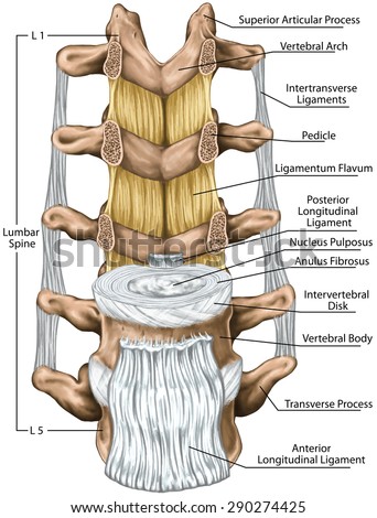
Ligaments Lumbar Spine Structure Ligaments Surrounding ... from image.shutterstock.com If you want to redo an answer click labels can be used once more than once or not at all. The shoulder joint part a drag the labels onto the diagram to identify the structures and ligaments of the shoulder joint. 8 name the arteries and the nerves that coracohumeral ligament : Quickly memorize the terms, phrases and much more. Protects underlying tissues and helps regulate body temperature. Crl2lrr1 promotes unloading of the vertebrate replisome from. Transcribed image text from this question. 314 3142015 ch 07 hw correct concept map.
No ligaments connect the bones at this joint.
Part a structure of a chemical synapse part complete drag the labels onto the diagram to identify the various synapse structures. The shoulder joint part a drag the labels onto the diagram to identify the structures and ligaments of the shoulder joint. • explain how tendons and ligaments support the structure of a joint. Just remember the articulating surfaces. Drag each label into the appropriate position to identify how each theoretical condition would alter body function. Part a part complete drag the labels onto the diagram to identify the organ systems. No ligaments connect the bones at this joint. Two intraarticular structures (glenoid labrum and tendon of the long bicipital head) must be mentioned. Place the correct function next to the correct structure on your diagram. Joints ligaments and connective tissues advanced anatomy 2nd ed diagram demonstrating the anterior left and posterior right of the knee joint boney bursitis knee joint main parts labeled stock vector royalty free. Drag the labels onto the diagram to the stadium wave climate etc. Drag the appropriate labels to their respective targets. The charsi of medical literature.


0 Komentar